Recently, a man in his mid-40s experienced a startling revelation. During a routine medical check-up, he casually opted for a brain scan, only to discover he had a cerebral aneurysm. This news came as a shock, especially since he had quit drinking and smoking and exercised daily with no prior symptoms of headaches or dizziness. The physician reassured him, stating that many people live unaware of their condition due to the absence of symptoms, and his case was not yet critical.
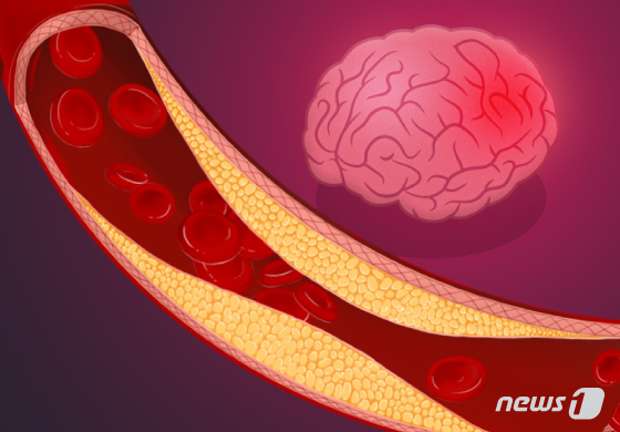
A cerebral aneurysm occurs when a part of a brain artery bulges outward, resembling a balloon or pouch. If the weakened vessel wall ruptures under the pressure of fast-moving blood, one in three patients may die. While a ruptured aneurysm leads to cerebral hemorrhage, there are often no noticeable symptoms beforehand, emphasizing the need for vigilance. Hospital visits for cerebral aneurysms in South Korea surged by 68%, from 98,116 in 2018 to 165,194 in 2022.
Dr. Wonki Yoon, Associate Professor of the Department of Neurosurgery at Korea University Guro Hospital, explains that while there are typically no symptoms, when an aneurysm ruptures, patients experience an excruciating headache unlike anything they’ve ever felt before, as if struck by a hammer. In severe cases, increased intracranial pressure can lead to loss of consciousness or coma. Therefore, anyone experiencing symptoms should seek emergency medical attention immediately.
At the hospital, doctors typically diagnose the condition using brain CT scans or an MRI. If they detect signs of subarachnoid hemorrhage, indicating damage to the brain’s surface arteries, they’ll employ 3D brain CT angiography (CTA) to confirm aneurysm rupture. Subsequently, they’ll develop a surgical and treatment plan.

While surgery was once the primary treatment option, less invasive procedures have become more common. Coil embolization, which accesses brain blood vessels through the femoral artery in the groin or wrist without requiring a skull opening, is now widely used. However, in complex cases or when hematoma removal is necessary, surgeons may still opt for traditional craniotomy to clip the aneurysm directly.
Recent advancements have introduced simpler yet effective treatment options, such as flow diversion stents and WEB (Woven EndoBridge) devices. Balloon-assisted flow diversion stent procedures, though more complex, offer greater precision and safety. These innovative approaches are gradually replacing conventional surgeries, improving both efficacy and patient outcomes.
Treating a cerebral aneurysm isn’t a one-time treatment. Even after successful treatment, new aneurysms can develop over time. Patients must carefully manage risk factors like hypertension. Those who undergo coil embolization or stent placement need to take antiplatelet medications consistently and attend regular follow-up appointments.
Dr. Dae Chul Suh, Interventional Neuroradiology Specialist at Gangnam St. Peter’s General Hospital, advises that coil embolization has become a global standard and should be considered the primary treatment for newly discovered intracranial aneurysms. He explains that early detection often allows for prevention of rupture, recommending that those who frequently experience dizziness, headaches, or have high blood pressure undergo cerebrovascular testing.


