
A groundbreaking study led by Professor Kim Cheol-hong from Pohang University of Science and Technology’s (POSTECH) Department of Electrical Engineering and Information Technology (IT) Convergence, in collaboration with Professor Choi Won-seok from Catholic University and researchers from Hebei Medical University in China, has unveiled a revolutionary technique for observing the placenta using cutting-edge medical imaging technology that combines light and sound.
POSTECH announced on Tuesday that this innovative research enables precise placental observation and early detection of pregnancy-related complications.
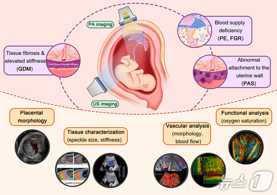
The placenta, a vital organ crucial for both maternal and fetal health, has long been challenging to study due to its temporary nature. To address this, the research team turned to photoacoustic imaging as a promising alternative to conventional ultrasound examinations.
Photoacoustic imaging harnesses ultrasound signals produced when hemoglobin in blood absorbs light, offering simultaneous visualization of vascular structures and oxygen levels. By integrating this method with traditional ultrasound’s deep tissue penetration capabilities, researchers can now comprehensively analyze placental structure, blood flow, and oxygen supply in a single examination.
Professor Kim Cheol-hong explained that its approach combines photoacoustic imaging with state-of-the-art ultrasound techniques to simultaneously assess placental blood flow and oxygen supply, overcoming the limitations of traditional ultrasound. This advancement is expected to significantly improve the early prediction and accuracy of major complications such as preeclampsia, fetal growth restriction, and premature birth.
The team’s findings have been published in the prestigious international journal Science Advances.


