The Korea Advanced Institute of Science and Technology (KAIST) said on the 21st that a research team led by Prof. Kang Ik-sung of its School of Electrical Engineering has developed a technology to precisely correct distortion in microscope images used to observe the interior of living organisms, in collaboration with a team led by Prof. Naji at the University of California, Berkeley in the United States.
The “two-photon fluorescence microscope” used in the study is a key instrument for observing deep within living biological tissue. However, as light bends and scatters while passing through thick tissue, images can become blurred, similar to how objects appear distorted underwater. This phenomenon is known as optical aberration.
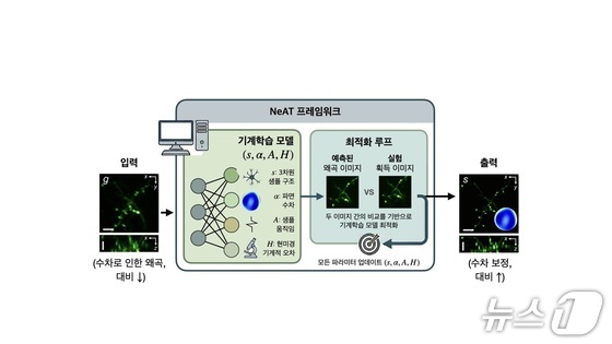
Previously, correcting such distortion required the addition of complex and expensive hardware equipment, such as wavefront sensors.
The research team developed an algorithm that calculates how light was distorted using only pre-captured image data and corrects it accordingly. Like restoring the original appearance from a blurred photograph, the method enables clear image reconstruction without additional equipment.
At the core of the technology is a machine learning algorithm based on a neural field model. The algorithm traces the process of light distortion during propagation, enabling an integrated correction of not only optical aberrations caused by biological tissue but also minute biological movements and mechanical errors of the microscope.
As a result, the team succeeded in stably obtaining high-resolution, high-contrast images from deep within biological tissue without the need for separate optical measurement or correction equipment.
The research team noted that the significance of the technology lies in overcoming the conventional limitation that “better images require more expensive equipment,” by solving the problem through a software-based approach.
Prof. Kang said, “This research opens a path to more accurately observe the interior of living organisms by combining optics and artificial intelligence technologies,” adding, “We plan to further develop this into an intelligent optical imaging system in which microscopes can autonomously find optimal images.”
The study was published in the international academic journal Nature Methods.


