

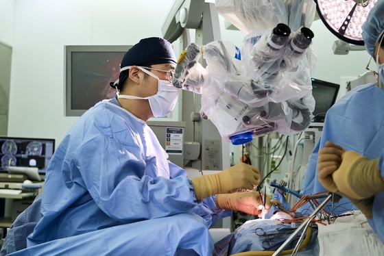
A woman in her 50s, Ms. Kim, who had long suffered from severe claustrophobia, unexpectedly discovered a left frontal lobe brain tumor during a routine health checkup. The tumor was dangerously close to the Broca’s area, responsible for speech, and the motor cortex controlling the tongue and face. This proximity posed a high risk of language impairment or motor paralysis post-surgery. Typically, an awake surgery would be performed to preserve language function, but Ms. Kim’s severe claustrophobia made this impossible.
In this critical moment, the neurosurgery team at Bundang Seoul National University Hospital, led by Professors Kim Jae-yong, Hwang Gi-hwan, and Kang Ho, offered a ray of hope. They employed two advanced techniques simultaneously: the cortico-cortical evoked potential (CCEP) method, which allows precise monitoring of language areas under general anesthesia, and the corticospinal radar, a motor nerve warning device.
Using CCEP, the surgical team pinpointed the exact location of the Broca’s area while Ms. Kim was under general anesthesia. They successfully removed the entire tumor while fully preserving her language function.
Professor Kang explained that it’s uncommon to discover a malignant glioma during a routine health checkup. However, the biopsy revealed a grade IV glioma, specifically a glioblastoma. This case exemplifies a successful proactive surgical intervention. Post-surgery, the patient experienced only minor speech difficulties, with her language function remaining largely intact. She has been living healthily for three years since the operation.
Maximal resection and safe preservation: The ultimate goal of glioma surgery
Gliomas, which originate within brain tissue and infiltrate normal brain matter, are among the most challenging brain tumors to treat. Removing these tumors requires a delicate balance to preserve vital brain functions.
The ultimate goal in glioma surgery is Maximal Safe Resection. This approach aims to remove as much of the tumor as possible while preventing neurological deterioration, ensuring patients retain their ability to walk, speak, and think normally post-surgery.
Professor Kang stated that maximizing tumor removal while ensuring patient safety may seem contradictory. However, it strives to find the optimal balance that allows for extensive tumor resection without worsening neurological symptoms.
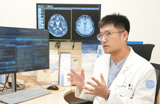
Preserving Language and Motor Functions During General Anesthesia: Cutting-Edge Neuro-Monitoring Technology
The glioma team at Bundang Seoul National University Hospital employs state-of-the-art neuro-monitoring techniques to precisely map brain areas and preserve their functions during surgery.
The corticospinal radar used in Ms. Kim’s case attaches a special electrical stimulator to a surgical suction device. This innovative tool sounds an alarm when approaching motor nerves, allowing real-time monitoring to minimize the risk of motor paralysis while safely removing tumors near critical areas. The glioma team uses this technique in about 60 to 80 surgeries annually, representing half of all glioma cases at the hospital.
Additionally, CCEP protects language functions in patients unsuitable for awake surgery. This method involves placing electrodes on the Broca’s and Wernicke’s areas, sending electrical signals to monitor language areas and nerve bundles in real-time. Currently, fewer than ten hospitals in South Korea actively use CCEP for brain tumor surgeries, with Bundang Seoul National University Hospital applying it in 15 to 20 complex cases yearly.
Professor Kang noted that the CCEP technique allows them to accurately locate the language cortex, which varies significantly among individuals, and monitor language nerve bundles during surgery. This advances them towards the goal of maximal safe resection.
The hospital has also fully implemented 5-ALA fluorescence-guided surgery. This technique involves administering a drug pre-surgery that causes malignant tumor tissue to glow pink in the operating room. Professor Kang emphasized that since pre-surgical magnetic resonance imaging (MRI) alone can’t definitively distinguish between grade II and III gliomas, it uses 5-ALA for nearly all grade II or higher glioma patients to enhance surgical precision.
From Diagnosis to Comprehensive Treatment: 3D Surgical Planning and Two Decades of Multidisciplinary Expertise
The glioma team’s strengths include thorough pre-surgical planning and a cohesive collaborative system. They create three-dimensional (3D) reconstructions of patients’ MRI images, allowing precise visualization of tumor shape, spread, and proximity to surrounding structures. This advanced planning, rarely utilized in clinical settings, enables careful review of surgical approaches before the operation.
Post-surgical care is equally robust. Glioma patients typically require tailored radiation and chemotherapy based on tissue biopsy results and genetic mutations.
Professor Kang explained that a multidisciplinary approach is crucial in brain tumor treatment. One neurosurgeon shouldn’t determine all aspects of care. The hospital pioneered a nationwide multidisciplinary brain tumor treatment system, fostering collaboration among neurosurgery, hematology-oncology, radiation oncology, radiology, and pathology to determine optimal treatment strategies.
Bundang Seoul National University Hospital established its brain tumor multidisciplinary team in 2006. Since 2013, they’ve implemented an outpatient system where patients can consult multiple specialists simultaneously. This collaborative approach covers everything from initial tumor resection planning to post-surgical chemo and radiation therapy scheduling. An integrated support center involving rehabilitation, mental health, and pain management specialists ensures comprehensive care for patients’ overall well-being.
Additionally, Professor Hwang Gi-hwan’s minimally invasive endoscopic surgeries for tumors around the pituitary gland and cavernous sinus showcase exceptional precision, further enhancing the team’s treatment capabilities.
Seek immediate medical attention for seizures or sudden language or motor impairments
Glioma symptoms vary based on tumor location and malignancy. Professor Kang advised that seizures are the most common symptom of low-grade gliomas. High-grade gliomas grow rapidly, potentially causing motor paralysis, language impairments, and cognitive decline within months. He urged anyone experiencing these symptoms to seek specialized medical care promptly.
Upon diagnosis, patients often first grapple with the possibility of a cure. Professor Kang explained that while some low-grade gliomas can be cured surgically, most are grade III or higher. These cases require post-surgical radiation and chemotherapy to delay disease progression as much as possible.
The glioma team at Bundang Seoul National University Hospital performs 100 to 150 glioma surgeries annually. They’re also advancing future medical research, including robotic surgery, epilepsy surgery integration, glioma organoid studies, genomic biomarker discovery, and artificial intelligence (AI) based imaging prediction models.
Professor Kim Jae-yong concluded that successful surgery is often the first crucial step in glioma treatment. While a brain tumor diagnosis can be overwhelming, it’s continuously improving the ability to achieve maximal safe resection through cutting-edge technology and multidisciplinary collaboration. It encourages patients to undergo necessary treatments with hope and determination to overcome this challenging illness.