Korean researchers have developed an artificial intelligence (AI) model that can identify whether a person currently has dementia and predict their future risk of developing the condition using only retinal fundus photographs.
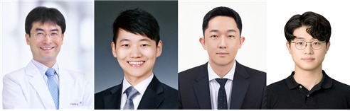
Seoul National University Hospital announced on Aug. 28 that a research team led by Professor Park Sang-min of Family Medicine, Dr. Jang Ju-young of Xymed, Assistant Professor Han Chang-ho of the Seoul National University College of Medicine Medical Big Data Research Center, and Dr. Kim Jae-won developed an AI model for screening and predicting dementia based on fundus images.
Dementia is diagnosed through a combination of clinical assessments, cognitive tests, and brain magnetic resonance imaging (MRI), but cost and accessibility limitations make large-scale screening difficult.
The retina, by contrast, is one of the few tissues in the human body where blood vessels and neural structures can be directly observed and is often described as a “window to the brain.” Previous studies have consistently reported changes in the retinal nerve fiber layer and vascular abnormalities in patients with Alzheimer’s disease.
The research team therefore used 108,008 fundus photographs from 36,322 people who underwent health checkups at Seoul National University Hospital between 2004 and 2016 to develop the AI model.
The researchers classified 1,001 people who were diagnosed with dementia within two years before or after their fundus photography as the dementia group and matched them with a control group without dementia at a 1:4 ratio. They then compared and evaluated 15 AI models combining five foundation models with three fine-tuning strategies.
The model that performed best was the “RETFound-MAE” model with partial fine-tuning, which had been pretrained on millions of fundus photographs.
The model achieved an AUROC of 0.750 for screening current dementia and a C-index of 0.812 for predicting future dementia. These results were higher than those of CAIDE, an established clinical dementia risk assessment tool, which recorded an AUROC of 0.624 and a C-index of 0.689.
Even after adjusting for existing risk factors such as age and sex, higher AI scores were significantly associated with greater current and future dementia risk. The researchers said this suggests that fundus photographs contain dementia-related information that cannot be explained solely by conventional risk factors.
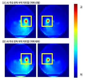
The researchers also applied explainable AI technology to examine how the AI reached its conclusions. The analysis showed that the AI paid the greatest attention to the optic disc and surrounding areas. Attention to the optic disc as a whole was 5.72 times higher than that given to surrounding regions, with the temporal region of the optic disc receiving the highest level of attention.
High attention was also observed in retinal regions previously associated with dementia, including the papillomacular bundle connecting the optic disc and macula. The researchers said the findings suggest that the AI may have assessed dementia risk using biologically meaningful retinal signals.
Professor Park said, “It is meaningful that dementia risk can potentially be assessed using fundus photographs taken during routine health checkups without requiring additional costly tests,” adding that he hopes the technology can become a useful supporting tool for dementia prevention and early intervention.
The study was published in the latest issue of the international journal npj Digital Medicine (IF 18.0).